© Dr. Tina MacGregor Reprinted in the ANVIL Magazine, March 1998 issue, with permission of Forge magazine. Solar punctures are one of the most common causes of lameness. They are usually the result of penetration of the solar surface of the foot by a sharp foreign body. As a result of this penetration, infection, usually by anaerobic bacteria, gains access to the hoof capsule and associated structures and an abscess develops. This article deals with the treatment of subsolar abscesses. This treatment can sometimes be an area of contention between farrier and veterinarian. When I was first given the title for this paper, it looked to me a bit like the title of a court case. So the case before us today is this: Is it necessary in the treatment of a subsolar abscess to make a big hole in the sole, or will a little hole suffice? Let us first define the terms: 'big' hole, and 'little' hole. These are subjective terms which do not actually describe what we are talking about. The question is, really, whether to remove all the underrun horn in a subsolar abscess or just make a drainage hole to allow removal of infected matter. So for 'big hole', read 'removal of underrun horn'; for 'little hole', read 'drainage hole without removal of underrun horn.' Before describing actual treatment, let me describe the events leading up to the formation of an abscess. Solar punctures in the horse are a relatively common injury. They may not always lead to infection of the internal structures of the foot with abscess formation. In my opinion, this is less likely to occur when the penetration is due to a nail prick during shoeing. It is more likely to occur when the penetration is the result of picking up a foreign body, such as a rusty nail, from the environment. The sequel to infection gaining access varies, depending on the site of penetration, the depth, the direction of penetration and the length of time the infection has been present.
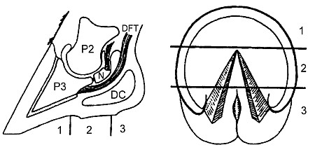
The foot can be divided into three zones: depending on which is penetrated, the infection can become trapped between the sole and the sole coriurm or the frog and the frog corium, without involvement of the corium itself or the underlying structures. This may be called a group 1 abscess and I shall return to this, as it is the area I wish to develop. If the penetration is deeper, it may result in the infection going into the corium itself or perhaps the distal phalanx, resulting in osteomyelitis. A penetration in zone 2 can have a severe outcome; there arc no hard structures to deflect the entry until navicular bone is reached. Deep penetrations in this region can lead to septic navicular bursitis, osteomyelitis of the navicular bone or possibly septic arthritis of the distal interphalangeal joint - very severe problems. In zone 3, deep penetration can lead to infection getting into the digital cushion or possibly the collateral cartilages. These deep infections are referred to as group 2 abscesses or infections. Their treatment often requires specialist surgical techniques, which I do not propose to discuss. It is important to be able to differentiate as quickly as possible between a group I and a group 2 abscess from the point of view of treatment and prognosis. I shall therefore concentrate on the group I abscesses and infections, which are far more common than the others.
Superficial penetration can result in the infection becoming trapped between the horn and the corium. The infection separates the horn from its corium, leaving the stratum germinativum covering the corium. Since the stratum germinativum is the horn-producing layer, it very quickly produces a layer of new horn, creating a cuticle to cover and protect the corium, and this has the effect of walling the corium off from the infective process. The infection is now trapped between two layers of horn. As a result of the bacterial infection, there is breakdown of horn and pus is produced. As the amount of pus increases in this relatively rigid horn hoof capsule, there is an increase in pressure inside the hoof capsule which affects sensitive structures and causes pain. The pain in a group I abscess, therefore, is a result of the increase in pressure. The effect is similar to that caused by hitting the fingernail with a hammer - the pain is a result of the pressure caused by a hematoma which forms under the nail. The pain caused by a group 2 abscess is due to the infection and destruction of the internal structures themselves. This difference is important when we come to consider the treatment of abscesses. It should also be remembered that these two groups are not mutually exclusive; a group I abscess can develop into a group 2 abscess. As a result of increased pressure in a limited space and the disruptive action of the bacteria, infection spreads. It takes the line of least resistance, between the horn and its corium, separating the two as it spreads. Abscesses under the sole tend to track backwards to the thinnest part of the sole, where they discharge. Abscesses under the wall tend to track upwards under the hoof wall, discharging at the hoof wall-skin junction. Abscesses under the frog tend to track backwards to the softest part which is the soft horn-skin junction under the bulb of the heels, where they discharge. It should always be remembered that if we find pus discharging from the foot here or we find evidence of a puncture wound at the toe, this may not be the full extent of the abscess. The infection may have spread quite a distance from the site of the actual puncture. What we see is simply the tip of the iceberg. A foot abscess produces clinically a sudden onset of lameness which may or may not have a history of foreign body penetration. In some cases there may be no foreign body present, and little evidence of penetration. That is particularly true of a wound in the frog region, where the tissue closes over the site of entry. The type of lameness depends on the area of the foot that is affected and the length of time the infection has been present; the lameness increases as the infection spreads. Where deeper structures of the foot are involved (group 2 abscesses), the lameness is more severe to begin, and very rapidly becomes worse. In most cases when moving the horse, try to place the foot so as to keep weight off the affected area - if the abscess is on the medial side, the horse tends to land on the lateral side. As the condition progresses, the horse will tend to bear weight only on the tip of the foot, regardless of where the abscess area is, and this can be misleading. The affected foot will tend to be hotter, and there will be a marked increase in the digital pulse. In the early stages, pressure with hoof testers, or percussion with a hammer may locate the area of the foot that is affected. But once the condition gets worse, wherever the foot is tested a pain reaction will result. If the horse is shod, the shoe should be carefully removed and each nail examined for evidence of pus or discharge. The foot should then be carefully examined with hoof testers, not only looking for a pain reaction but also for softening in an area, or perhaps a discharge. The foot should then be thoroughly searched by shaving the surface area of the sole, white line and frog. At this stage, we are looking for anything that might suggest a site of penetration or the track of the foreign body or evidence of bruising or abnormal softening of the horn, which would require further investigation.
Treatment If the horn is very hard and difficult to pare and all the clinical findings suggest there is a subsolar abscess, it may be worth considering soaking the foot in water overnight, allowing the horn to absorb water and become softer. Salt should not be added to the water as this makes it hypertonic - resulting in water being drawn out of the foot, making the hoof horn harder. Similarly, poulticing with hypertonic substances should be avoided, as it tends to harden the foot. Once the abscess pocket is reached, pus will drain from the hole. In the case of a group I abscess, the pus is usually colored gray, black or brown, the color being due to bacterial breakdown of horn. The pus from a group 2 abscess is usually much paler - the usual pus color, yellow or creamy. It is important to note the color, as the paler pus may be the first indication that the deeper structures of the foot are involved. Having established drainage, the pressure is reduced on the sensitive structures and the horse may quickly show a marked improvement in lameness. The treatment, however, is far from complete. Bacteria remain inside the foot. All the infected material needs to be removed from under the sole to prevent spread of infection, or reinfection. This is where the decision must be made, to make a big hole or a little hole. Or, in other words, remove all underrun horn or make an access hole for drainage without removal of underrun horn.
Two Schools of Thought The other school of thought, by contrast, believes that all of the underrun horn should be removed, regardless of the extent of the area involved. In some cases, particularly when the treatment is done at an early stage, the question is academic, as all the underrun horn may be removed by the creation of the access hole, if the abscess pocket is very small. If the abscess is advanced, however, the underrun area may be quite extensive. That is when the question arises, which approach should be taken. The advantages of the small hole approach, with flushing, are that there is less disruption of the hoof capsule, which makes owners happier. The hole will also tend to heal over more quickly than if a larger area is exposed and there is less chance of the foot picking up external contamination. Some people also believe that the separated corium is protected from injury this way, the underrun horn acting as a cover. But in most cases, the corium will already have formed its own protective layer of horn, which tends to remain soft unless exposed to air. The disadvantages of this form of treatment are, first, that not all of the infected tissue may be removed; and if infected tissue remains inside the foot, reinfection will quickly occur. Although flushing through the small hole will remove bacteria, there is also a risk that it will force bacteria further into the foot, particularly if it is done under pressure. In that case, the bacteria will become inaccessible. Further, by not removing the underrun horn, there is a strong chance that the anaerobic environment will allow many of the bacteria that caused the infection in the first place to persist. A small hole also prevents visual examination of the corium to see whether it is intact and healthy. And because a small hole tends to heal over more quickly, any developments or further deterioration such as spread of infection into deeper structures will go undetected until further clinical signs develop. Occasionally, if too small a hole is made, because of the pressure inside the corium at the base of the hole, it may prolapse through the hole and become trapped or pinched. This is extremely painful and can make the horse more lame than it was before treatment started. Leaving the underrun horn also creates a dead space beneath the corium, which produces a new sole. There are thus two soles, old and new, with a dead space between; this can become filled with debris which puts pressure on the new, soft sole and can keep the horse lame.
Method of Choice The healing process can be readily observed to see if all the underrun horn has been removed and that there is no further discharge which would indicate that a group I abscess is developing into a group 2 abscess. The progress of the newly developing horn can also be observed, to ensure that it becomes firm, dry and hard. Another advantage is that pinching of the corium is unlikely, as is the development of a double sole impacted with debris, leading to persistent lameness. This is not to say that there are no disadvantages to the 'big hole' approach. First, it may take longer to heal over which is often the main concern of the owner. And exposure of a large area of corium may create the risk of damage during or after the paring process. However, although removal of a large amount of horn may look bad at first, in actual fact, paring the foot right at the start and getting to the root of the problem immediately may actually speed up the heating process in the long run. Damage to the exposed corium may be avoided by careful paring with a blunt probe to guide and assess the extent of the underrun area and ensuring that the foot is protected and supported after the area is exposed. A dry dressing is best for this; if an antiseptic solution or antibiotic spray is used, it is more difficult to assess the progress of healing whether the corium is becoming dry or still discharging - and masks the nature and color of any discharge. The proprietary nappy, Pampers, (the boy's type), fixed to the foot with adhesive tape, has been found to be an effective dry dressing. The dressing should be changed, preferably daily, until there is no longer any discharge and the horn over the corium is dry and firm to the touch. In my opinion, systemic antibiotics are of little value in the treatment of subsolar abscesses, as the antibiotic cannot penetrate effectively to the affected area, which is isolated between two layers of horn. That is the case for and against the big and small hole approach. You must make up your own mind as to the approach you yourself favor.
Roles of Farrier and Veterinarian Dr. Tina MacGregor, BVMS, MRCVS, is an international authority on the equine foot. This article is adapted from a presentation she gave at the NAFBAE/BSAVA seminar. Return to the Farrier Articles listing page. Return to the ANVIL Online Table of Contents for March, 1998.
|