
© Yehuda Avisar, DVM
published in ANVIL Magazine, December, 1995
INTRODUCTION
Contracted heels in the horse is characterized by a shift of the hoof wall resulting in the narrowing of the foot. Primary cases of contracted heels result from unbalanced feet, either long toe/low heels (LTLH) or overgrown hooves. Trimming the feet and correcting the hoof angle result in expansion of the heels. Secondary contracted heels are caused by lameness and disuse of the limb, resulting in hoof atrophy.
Problems associated with contracted heels include disruption of the shock-absorption mechanism of the foot (Rooney 1974). The contracted hoof wall presses internal structures, a condition termed hoof bound (Rick 1907). Crevices and grooves formed by the contracting hoof creates a favorable environment for thrush.
FACTORS LEADING TO LONG TOE/LOW HEEL
Some horses' hooves tend to grow the toe at a faster rate than the heels.
Expansion and contraction of the heels during motion increases their wear relative to the toe.
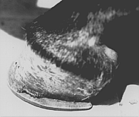
(The photo to the left shows typical underrun heels. The heels are dropped, and the bulb of the heels are prominent. The condition can be caused by a horseshoe that is too small, as can be seen here.)
Many horses have naturally underrun, weak heels. The heels are lowered more than necessary, creating a broken foot axis.
CLINICAL APPEARANCE
Initial cases of primary contracted heels are not lame. Any foot can become affected, but the condition is more common in the forelegs. The hoof appearance is similar to the shape of a mule's hoof. In some cases the entire hoof is contracted, the sole has more concavity and the frog atrophies. Some horses develop heel bulb contraction.
RULE OF HOOF MOISTURE
Moisture content in the hoof is maintained by circulation and is influenced by the environment. Dynamic movement of the hoof during exercise increases the circulation within the foot (Butzow 1961). The elasticity of the hoof is dependent on the moisture content of its keratinized structures, and lack of moisture reduces resiliency of the hoof, affecting shock absorption (Emery et al 1977).
Lack of exercise, prolonged stabling in dry stalls, hoof dressings, sand and urine tend to dry the hoof. Lambert 1968 demonstrated rapid loss of moisture and contraction of the heels following regular hoof trimming.
SHOCK ABSORPTION
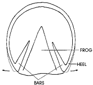
(A solar view of the hoof. The schematic representation is of the area involved in shock absorbtion. The dotted line is the area occuppied by the quarters and heels during expansion of the hoof wall.)
The caudal part of the hoof wall functions like a spring, spreading apart as the foot contacts the ground, absorbing the energy of impact. The outward movement of the heels and quarters is permitted by the flexible frog, the bars support the hoof wall and assist in their return to a non-weight-bearing position as weight is removed. Proper function of this spring depends on resiliency of the keratinized structures of the hoof. The dynamic movement of the hoof wall creates a pumping action that increases circulation within the foot (Lambert 1966).
HYPOTHESIS FOR PRIMARY CONTRACTED HEELS
Many authors (Emery et al 1977, Dollars 1898, Hays 1968, Lungwitz 1966 and Stashak 1987) indicate concurrency of LTLH and contracted heels. Stashak 1987 recommends balancing the feet for treatment of contracted heels, indicating that some cases can be resolved after a single trimming. The same observation was made by this author. Increasing hoof angle by 3 to 5 degrees and balancing the foot with the same level of activity resulted in expansion of the heels within a few weeks. This observation led to the following hypotheses regarding the development of primary contracted heels in the horse: lowering the heels or LTLH increases tension on the deep digital flexor tendon (DDFT). The tension pulls the deep digital flexor muscle (DDFM), causing it to stretch. The DDFM opposes the stretch by contracting. The contraction of the muscle, caused by a stretch reflex involving nerve fibers in the muscle, signal when the muscle is suddenly stretched; the static stretch reflex continues to cause muscle contraction as long as the muscle is maintained at excessive length (Guyton 1986). When the LTLH condition is maintained, the muscle will continue to oppose that force. The contraction of the DDFM pulls the caudal aspect of the foot, and weight distribution to the heels is decreased.
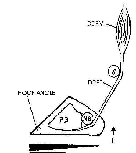
(Development of contracted heels depicted diagrammatically. Contraction of the deep digital flexor muscle (DDFM) lifts the caudal hoof, decreasing normal weight placement to the heels (arrow). The black triangle represents the relative weight distribution to the bottom of the hoof. The navicular bone (NB) and the sesamoid bones (S) normally function as pulleys, for the deep digital flexor tendon (DDFT) and may actually cause lifting of the heels.)
During weight bearing, the heels don't reach maximal expansion; circulation within the foot decreases and the keratinized structures lose moisture and contract. Contraction is more rapid at the heels since the bulbs of the heels are less resistant to the contracting forces. Contraction of the heels develops in two planes, narrowing and curling of the hoof wall.
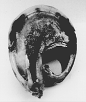
(To the left is shown a hoof one hour after being detached from a laminitic case. The left bar and a circular section of the sole were removed, decreasing the resistance to narrowing and curling of the hoof wall.)
When contraction is severe, the bulbs of the heels begin to contract. The caudal hoof wall becomes more upright and loses the spring action of the heels. Impact is being transmitted to bones and ligaments, increasing the chance for limb injury.
TREATMENT
Most treatment regimens for contracted heels are aimed at treating the symptoms and not the cause (Lambert 1966). Treatment principles include trimming and balancing the feet and assisting heel expansion. Primary contracted heels have to be differentiated from secondary contracted heels, caused by a painful condition in the foot or disuse of the limb. Berns 1918 described the long-term effect of contracted heels on a group of horses. The clinical signs were similar to navicular disease, but after trimming and expanding the hooves, the horses became sound.
Numerous treatment methods appear in the literature. Some can aid in the expansion of the heels, while others can be harmful. Methods include: lowering the heels to increase frog pressure (Simpson 1968); special steel shoes for contracted heels (Russel 1882); springs applied against the bars and special spreading devices (Lungwitz 1966); mushroom shoes, tips, bar shoes and pads; beveled horseshoes (The Horseshoer1966); grooving or rasping the hoof wall (Stashak 1987); Jenny et al 1965 recommend the use of acrylics to treat contracted heels.
Increasing frog pressure has no effect on heel expansion, and lowering the heels can lead to further contraction. Tip shoes are more natural to the hoof but tend to concentrate pressure on the toe, separating the hoof wall at the white line. Beveling the horseshoe hoof surface can aid in heel expansion, but rasping and grooving the hoof wall can cause loss of moisture from the hoof. Spreading devices and springs should be used with caution or avoided.
Overgrown hooves should be trimmed short, and the horse exercised daily. Some animals may have sore feet following trimming, but will improve gradually. Berns 1918 recommends pine tar and oakum held by a leather pad. Leaving the horse barefoot and exercising it is another method. Acrylics can be used to reconstruct underrun heels; a new product that uses rubber particles that are glued to the bottom of the hoof wall may aid in heel reconstruction (Ferguson 1994).
Horses with LTLH should be trimmed every 3-4 weeks to maintain a constant hoof angle. Rick 1907 recommends trimming the feet to establish a 53 degree for the forelegs and a 58 degree angle for the hind legs. These figures are higher and more correct when compared to figures measured randomly on modern-day horses. Breed, function, conformation, gait and individual pattern of hoof growth has to be considered when balancing the horse's hooves.
Increasing the hoof angle can resolve the condition for some of the cases, sometimes in one treatment (Stashak 1987). Once the heels expand, the groove formed between the bulbs of the heels opens.
Hoof moisture can be increased by dampening the paddock with water or applying a commercial poultice, especially following trimming. Hooves afflicted with thrush are trimmed until healthy tissue is reached and pine tar is applied to the exposed areas.
If shoeing is required, the horseshoe nails are driven anterior to the midquarters, and the horseshoe is fit gradually wider at the quarters and heels, extending about two centimeters backwards to support the heels.
(An excellent example of the relationship of the horseshoe to the contracted hoof. The hoof surface of the horseshoe was beveled to encourage heel expansion.)
A good contact between the hoof and horseshoe is important - this will allow uninterrupted expansion and contraction of the heels during motion.
REFERENCES
Berns G.H. (1918) Lameness of obscure origin and some of its causes. 55th Annual Meeting, AVMA, 217-222.
Butzow R.F. (1961) Anatomy and care of the equine foot. Illinois Veterinarian 4 (4):98-103.
Emery L., Miller J., Van Hoosen N. (1977) Natural state and function of the hoof. In: Horseshoeing Theory and Hoof Care, 1st edition. Lea and Febiger, Philadelphia. p 78.
Ferguson J. (1994) University of Florida, College of Veterinary Medicine, Large Animal Clinical Sciences. Personal communication.
Guyton A.C. (1986) The stretch reflex. In: Textbook of Medical Physiology, 7th ed. W.B. Saunders. pp 609-611.
Jenny J., Evans L.H., Raker C.W. (1965) Hoof repair with plastics. JAVMA 147, (12)1340-1345.
Lambert F. (1966) The role of moisture in the physiology of the hoof of the harness horse. VM/SAC 61(4)342-347.
Lambert F. (1968) An experiment demonstrating rapid contraction of a standardbred horse hooves from moisture loss during flooring. Vet. Med. Small Animal Clinician 63, 878-881.
Lungwitz A. (1966) Defects of the hoof. In: A Textbook of Horseshoeing, 11th edition. Trans. J.W. Adams. Oregon State University Press, Corvallis, OR. pp 185-190.
Rick G.E. (1907) Chapters V and VI. In: Rick's New Artistic Horseshoeing, 1st edition. The Commercial Printing Co. Akron, Ohio. pp 86-87 and 101-102.
Rooney J.R. (1974) Foreleg lameness. In: The Lame Horse, 1st edition. A.S. Barnes and Co., Inc., Cranbury, New Jersey. pp 136-137.
Russel W. (1882) Diseases of the foot. In: Scientific Horseshoeing, 1st edition. Robert Clark and Co., Cincinnati. pp 69-72.
Simpson J.F. (1968) The theory of shoeing and balancing. In: Care and Training of the Trotter and Pacer, 1st edition. Ed. J.C. Harrison. The United States Trotting Association, Columbus. pp 369-371.
Stashak T.S. (1987) Methods of corrective trimming and shoeing. In: Adams' Lameness in Horses, 4th edition. Lea and Febiger, Philadelphia. pp 825-825
U.S. War Department (1966) Normal, special and corrective shoeing. In: The Horseshoer. California Polytechnic State University, San Luis Obispo, CA. pp 70-73. ¨
Dr. Avisar is a practicing equine veterinarian in Neoth, Golan, Israel. For 17 years before becoming a veterinarian, he worked as a farrier, including seven years with UC Davis Veterinary School farrier, Charles A. Heumphreus.
Return to Veterinary/Technical Articles
Return to the Veterinary/Technical Articles page