Published in the October 1999 Issue of Anvil Magazine 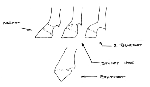 Fig 1 Drawings
by Lungwitz (1910). Fig 1 Drawings
by Lungwitz (1910).
So-called ōclubfootö has long been a vexing problem for horsemen, veterinarians, and farriers. The term clubfoot is a misnomer for the condition in the horse and correctly refers only to a congenital anomaly of the human foot. Lungwitz (1910) properly defined and described the condition for the horse. There are three forms: 1. Stumpy hoof. (Figs. 1, 2) This is bockhuf, goat hoof, in German, but will be translated here as stumpy hoof. The hoof angle is greater than normal, 60-90 degrees. The pastern is short or appears so and is usually more or less in line with the hoof axis as seen from the side. 2. Bearfoot. (Fig.1) This is ba¨renfuss in German. The hoof angle is greater than normal, as above, but the pastern appears longer and has a more sloping angle, ō...broken strongly forward at the coronet.ö Lungwitz(1913). 3. Stiltfoot. (Fig.1) This is stelzfuss in German. It is the extreme or final case of 1. and 2. with the foot supported only at the toe. The major distinguishing characteristic of stumpy hoof or bearfoot — that which compels the attention of the observer — is the consistently greater angle of one hoof as compared to the other. The basic abnormality in all three cases is shortening of the deep flexor tendon with or without prior or accompanying shortening of the superficial flexor and suspensory tendons. The term ōcontracture,ö usually used to describe these conditions, is a misnomer and will not be used here. Tendons do not and cannot contract, either normally or pathologically. Apparent contracture, either bilateral or unilateral, is shortening of a tendon or tendons towards the normal rest length. The rest length is that length the tendon assumes when not under tension. While the primary purpose of this paper is to discuss shortening of the deep flexor tendon, it is necessary and appropriate to summarize the pathogenesis of yearling tendon shortening as well. (The more mathematical material has been relegated to the Appendix, but that does not mean it should be ignored.) It must be emphasized that much of what is presented here is hypothetical and based on gleaned experience and mechanical principles. There is no experimental data available directly relevant to the questions examined, and it is difficult to conceive of experiments which could reasonably be done.
Yearling Bilateral Tendon Shortening The pathogenesis of so-called yearling contracture has been presented elsewhere (Rooney 1984, 1997). The mechanics are summarized in the Appendix. To review: with rapid weight gain, the immature hoof is forced into a low heel, small hoof angle (as measured at the toe). This change of hoof angle increases the tension on the deep flexor tendon by rotation of the coffin joint. The subsequent increased tension in the deep flexor at the fetlock joint is equilibrated by decreased tension in the suspensory and superficial flexor tendons. As the tension decreases in these two tendons, they shorten; and, in doing so, it moves the digit into the more upright position characteristic of yearling tendon shortening. With continuing shortening of the superficial flexor and suspensory, the deep flexor will also shorten. While this is occurring, the increasing body weight (weight gain), continues to force the immature hoof into a low hoof angle.
Fig. 2 Photograph of stumpy foot of the left foot. These changes are taking place while the tendons are still maturing, before cross-linking of the collagen fibers has been completed. Permanent, irreparable deformity may be the result if the shortening process is prolonged before corrective measures are taken; that is, if the tendons become fully mature and cross-linked while in the shortened state. It is now generally recognized that many cases of yearling tendon shortening will correct themselves if weight gain is restricted or stopped before the process has proceeded to ōknuckling-overö (stiltfoot). The reason should be apparent from the above discussion. The cause of bilateral tendon shortening before a year of age is due to the fact that the immature hoof is unable to maintain a steady hoof angle while there is a rapid gain in weight. There are, therefore, both genetic and environmental factors. The genetic factor is the rate of maturation of the hoof wall; the environmental factor is rapid weight gain. The latter, of course, also has a genetic component. Unilateral or Deep Flexor (Stumpy Hoof/Club Foot) Shortening These clinical observations are largely anecdotal from personal observation and discussions with farriers and veterinarians. 1. The right fore foot is more often affected, about 70-80%. Both fore feet are rarely affected. Since this condition develops in an insidious manner over time, the age of onset has been difficult to determine, either for specific cases or in general. Most cases probably begin during the first year of life while others may not appear until the horse is an adult. This will be further addressed below. 2. The carpus of the affected leg is higher than the contralateral carpus. 3. The scapulohumeral angle may be smaller on the affected side. 4. The dorsal wall of the hoof may be dished. 5. The affected leg almost always stands back. 6. There may be atrophy of the musculature of the affected foreleg. 7. The breed incidence is not known. It has been reported in many (but not all) breeds, and anecdotal evidence has suggested there may be predilection for Arabians and Arabian crosses. Lungwitz (1910) categorically states that stumpy/bear foot is seen in all breeds and in both fore and hind legs. I have not seen the condition in the hind legs. There are four scenarios which can be associated with the pathogenesis of stumpy/club foot. The final pathological pathway is always abnormal shortening of the deep flexor tendon, but this final pathway may be reached by any of four ōfeederö routes or a combination thereof.
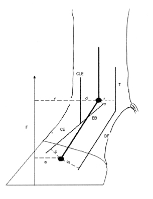
Fig.3 The moment applicable in the study of the mechanics of flexor and suspensory tendon shortening. See the text for explanation. 1. Pain. If the animal is unwilling to bear weight on a painful forefoot over a period of time, the F term, the ground reaction force in equation [1] (Fig. 3, Appendix) is too small or absent. The CEc moment, that moment produced by the common extensor tendon and the extensor branches of the suspensory, is not large enough to equilibrate the equation and, so, DF decreases — the tension in the deep flexor tendon decreases and that tendon shortens. When the deep flexor tendon shortens, the heels and quarters are lifted from the shoe or surface or, at least, bear less weight than the toe and sidewalls of the hoof. This allows the heels and quarters to grow out, increasing the hoof angle. With increase in hoof angle, the animal will tend to ōstand backö (Fig. 4). This process can be visualized in Fig. 5, which shows via the diagram how the increase of hoof angle causes the leg to move back under the body. Such a scenario is likely to lead to permanent stumpy foot if the process begins during the first year of life while the tendons are still growing and maturing. (See the discussion of collagen cross-linking with the discussion on yearling tendon shortening, above.) Additional factors come into play with the ōstanding backö position of the leg, although the effects to be described are not great. With the leg ōstanding back,ö the coffin and fetlock joints are both dorsiflexed, and there is less tension in the common extensor tendon and the extensor branches of the suspensory tendon. (Rooney, 1969) This means there is less resistance to the tension in the deep flexor tendon, and the deep flexor tendon shortens. The ōstanding backö position, then, predisposes to shortening of the deep flexor tendon which in turn allows the hoof angle to increase. Once the process has progressed far enough, the coffin joint is moved from dorsiflexion to palmarflexion by the shortening of the deep flexor, and the lesser tension in the common extensor and extensor branches is dependent now only upon the dorsiflexion of the fetlock joint. As the reciprocal process of hoof angle increases and deep flexor shortening continues, the affected leg is less stable, and the horse may bear even less weight on that leg — allowing the deep flexor to shorten even more. The changes in conformation of the affected leg, listed previously, including muscle atrophy and changes of shoulder joint angle, can be ascribed to the ōstanding-backö position and less weight bearing by the affected leg.
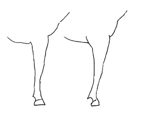
Fig 4. The usual, normal standing position of the foreleg to the left and "standing-back" position to the right. 2. Abnormal Wear Owen (1975) observed the development of stumpy hoof conformation in foals confined for long periods to horse boxes during bad weather. The foals obsessively pawed the ground, wearing away the toe and so producing the high hoof angle which allowed shortening of the deep flexor tendon. Again, though not specifically discussed by Owen, these foals would stand back on the large angle hoof and exacerbate the condition as described above. Lungwitz (1910) mentions lack of attention to trimming the feet of foals running barefoot as a causative factor. This is akin to the OwenÆs cases. 3. Environmental First, we will consider that the animal is entirely normal before the process to be described begins, so that only environmental factors are operating. The foot about to undergo deep flexor shortening has too small a hoof angle and the animal is gaining weight. This is basically the same condition as for yearling tendon shortening, with the important exception that the hoof is more mature and able to bear increasing weight without deforming to the smaller hoof angle characteristic of yearling tendon shortening. The shortening deep flexor, in this case, tends to lift the quarters and heels from the surface (or the shoe). This, in turn, allows the heels to grow because of less wear, and the angle of the hoof increases — the opposite of the yearling situation. Increasing hoof angle means that the coffin joint is rotating, so that tension in the deep flexor continues to decrease. That is, the deep flexor continues to shorten. The conditions which started this process — low hoof angle and increasing weight — are now changing to high hoof angle with less weight being borne by the affected leg. As the hoof angle increases, the leg tends to move to the standing back posture, which exacerbates deep flexor shortening. It is to be reiterated that increasing weight (the ground reaction force, F, of the Appendix, Fig.3) is an important factor in the early stages of this type of tendon shortening sequence, while decreased weight bearing is important in the latter stages.
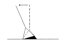
Fig. 5 If the heel grows out (the black portion) the normal position of the leg, shown as the dotted lines, shifts to the new, standing back position, the solid lines.The head of the horse is to the left 4. Inherited Stumpy/Bear Foot The stumpy/bear foot conformation can be an inherited characteristic per se. There appears to be no hard data in the literature, but the anecdotal experience of farriers and veterinarians suggest that there is often a genetic component: affected mares and stallions tending to have affected offspring. Lungwitz (1910) considered both bear foot and stumpy foot to be congenital. While it appears probable that many stumpy feet are of congenital/genetic origin, this may not become manifest until years later if the affected foot has been carefully trimmed and shod in the interim. By the time the condition becomes obvious, the early stages — the conformation at birth or pain or rapid growth — have long since changed and are unlikely to be remembered. Consider the foal born with a bear or stumpy foot. The stumpy leg will tend to stand back. Once again, to repeat, in this position the coffin joint is dorsiflexing, which tends to loosen the common extensor and extensor branches of the suspensory, while the fetlock is dorsiflexed more than in the normal standing position. This allows the extensor branches of the suspensory to loosen. The result is decreased tension in and shortening of the common extensor and extensor branches. For equilibrium at the coffin joint, the tension in the deep flexor must decrease, and the deep flexor shortens. The hoof angle will gradually increase because of less heel wear with consequent shortening of the deep flexor tendon as already described. The loosened common extensor and extensor branches will, of course, be tightened (increased tension) as the hoof angle increases, but this is countered by the shortening of the deep flexor occasioned by the increasing hoof angle. I have not encountered reports of stumpy foot in Standardbred horses. Yearling tendon shortening does occur in Standardbreds. Since Standardbreds share genetic heritage with a number of other breeds which do have stumpy foot, one can speculate that it is the singular attention and care given to the feet of many of these horses which could be the protective and/or preventive factor. Further to that point, anecdotal evidence suggests that stumpy foot is more common in horses used for purposes other than racing. That could be related to the more frequent trimming and shoeing of racing horses, both Standardbred and Thoroughbred. Racing horses are exercised and examined frequently and kept in usually thinner condition than is typical of weekend and show horses. Hood et al 1997, showed with force plate studies that horses over time tend to bear weight preferentially on the left or the right side and tend to shift weight on the diagonal. They suggested this could be related, presumably, to genetically determined ōhandedness.ö Horses predisposed to stumpy foot of the right fore, for example, could be bearing more weight for more of the time on the right fore and the left hind since weight on the leg is a factor in the early stages of the development of stumpy foot. Appendix If the hoof angle is decreased as by cutting down the heels, the coffin bone is rotated clockwise (with the horse facing left), increasing tension in the deep flexor tendon. This increase of tension will tend to push the fetlock up and forward, the pastern moving to a more upright, vertical position. The moment equation for the coffin joint is, (Fig.3): DFb-CEc-Fa = 0 1. where DF is the linear tensile force in the deep flexor tendon, CE is the linear tensile force in the common extensor tendon and extensor branches of the suspensory tendon, and F is the ground reaction force. a, b, and c are the moment arms, the perpendicular distances from the respective linear forces to the center of rotation in the distal end of the middle phalanx. The moment equation for the fetlock joint is (Fig.3) given in simplified form sufficient for present purposes: Tr-Ff-CLEd Ò EBe = 0 2. where T is the sum of the tensile forces in the superficial and deep flexor tendons and the suspensory tendon. r is the average moment arm of these three tendons, acting around the center of rotation in the distal end of metacarpal 3. F is the ground reaction force and f its moment arm around the that same center of rotation. CLEd is the moment around the fetlock produced by the common extensor and lateral extensor tendons. EBe is the moment produced at the fetlock by the extensor branches of the suspensory tendon. If the fetlock dorsiflexes without movement of the coffin joint, the moment EBe is negative (-). When the coffin palmar flexes, followed or accompanied by dorsiflexion of the fetlock, the extensor branches slide distally, so that the moment EBe becomes positive (+). When the angle of the hoof decreases, for whatever reason, the moment arm, a, at the coffin joint increases and the moment equation is not in equilibrium. In order to regain equilibrium, DF, the tensile force, in the deep flexor tendon, increases. That entails an increase of T, the tensile force acting at the fetlock since DF is part of T. Equilibrium is then lost at the fetlock and is regained by a decrease of tension of the superficial flexor and suspensory. As the tension decreases in these two tendons, they shorten, and the pastern becomes more upright. The moment arm, f, of the force, F, decreases as the pastern becomes more upright, and so the moment Ff decreases, allowing for additional decrease of T and the pastern becomes more upright as the deep flexor, superficial flexor, and suspen-sory all shorten. Bibliography 1. Hood, D M, Hunter, J F, Beltz, W D, Taylor, B E, Beckham A S and Pierce (1997) J R Digital Loading Patterns in the Normal Standing Horse. Proceedings of the Hoof Project, 37-43. 2. Lungwitz, A. Trans. Adams, J.W. (1913) Horseshoeing. Facsimile edition, Oregon State University Press, Corvallis. 3. Lungwitz, M. (1910) Leisering u. Hartmann, Der Fuss des Pferdes. 11th Ed., Schaper, Hannover. 4. Owen J M 1975 Abnormal flexion of the corono-pedal joint of ōcontracted tendonsö in unweaned foals. Equine Veterinary Journal 7:40-45.
|